 Most congenital disease are of unknown cause. This disease is not an exception.
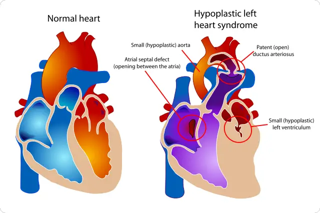
Hypoplastic Left Heart Syndrome or HLHS is a disease that manifests immediately after the birth of an infant.
Fact #1: Elaboration of the medical terms
The term hypoplasia refers to the congenital condition of a tissue or organ. Hypo means small. Therefore, in this case, the left atrium and left ventricle is underdeveloped, including the mitral and aortic valves are small too.
Fact #2: How does a normal heart work and the effects of a small left heart.
Most of the deoxygenated blood in the body will return back to the heart though the inferior and superior vena cava. It will be received by the first chamber: right atrium, then the tricuspid valve will open so that the right ventricle can pump this blood to the lungs through the pulmonary artery. Gas exchange will occur and the oxygenated blood will return to the heart though the pulmonary vein. Left atrium will receive the oxygenated blood then the mitral valve will open to let the blood to flow to the last chamber: left ventricle. The oxygenated blood will be forced out of the heart and delivered to the rest of the tissue through the aorta.
The left side of the heart pumps with a greater pressure because it distributes the oxygenated blood to the whole body. Imagine if its size is smaller, cardiac muscles could not withstand the pressure of pumping the blood, less oxygen results to tissue hypoxia, and loss of function of other systems of the body without the air that gives them life.
Fact #3: For these children being more abnormal is a higher chance of survival
Lungs of fetus do not work inside the mother’s womb. Oxygen is supplied by the umbilical vein and waste products flows to the umbilical artery. The baby receives the oxygenated blood through the right atrium, but it bypasses the right ventricle because of the hole between these two opposite chambers: foramen ovale. Some of the blood that goes to the right ventricle will go to the pulmonary artery but flow straight to the aorta because of another hole that connects these arteries: ductus arteriosus. Therefore during the whole gestational weeks, the lungs are nonfunctioning. Problems will only arise after the baby’s first breath.
Normally, these two shunts: ductus arteriosus and foramen ovale closes after birth. Congestive heart failure may arise if the shunts would remain open. However, for babies with HLHS, closure of the shunts are prevented. If ductus arteriosus and foramen ovale remained open for a longer time, it would be more favorable for them. The right ventricle will do all the work of pumping the blood to be oxygenated in the lungs and distribute it to the rest of the body because the left side of the heart does not work well.
Once these holes close, the weak left side of the heart will function and the babies will be at the verge of their death.
Fact #4: Signs and Symptoms
Abnormalities of these babies arise from their body’s oxygen insufficiency. They are actually termed as “blue babies” because of their cyanotic skin color. You have observed that veins are grayish blue because they are poorly oxgenated. All of the blood vessels in their body is oxygen deficient that is why their skin color is like that.
Other manifestations are difficulty of breathing, cold hands and feet, dizziness and lack of physical activity.
Fact#4: Diagnosis
Abnormality could be detected during the pregnant mother’s ultrasound. Other means of diagnosis is echocardiogram. This is a detailed imaging of the structure of the heart through a transducer that converts the soundwaves into detailed imaging of the heart.
Fact #5: Management
Babies with HLHS are misfortunate because they feel too tired while eating. A special high calorie is prescribed and they are fed through a special tube.
Different procedures are deviced to lengthen the patient’s survival. Norwood procedure is done after two weeks of birth. All arteries are connected to the right ventricle so the fragile left ventricle will not be allowed to work anymore. After 4-6 months of age, the pulmonary artery and superior vena cava will be connected through the Bi-Directional Glenn Shunt procedure. This is to lessen the burden of the right ventricle. The final surgery, Fontan procedure connects the inferior vena cava and pulmonary artery so that all of the deoxygenated blood will no longer pass through the right chambers.


Leave a Reply